Total Care for Women provides obsterical and gynecologic Ultrasound services on site!
Links on this page
Saline Sonohysterography (SIS)
What is Ultrasound Imaging?
Ultrasound developed in the mid 1970's from simple measurements of body parts to now 3 D computer modeling of body parts. The science is still high frequency sounds waves theat are transmitted through tissues and reflected back to the receiver. Body tissues reflect the minute wave energy back from each tissue interface. With modern computer technology, the direction, intensity and time delays all contribute to form an image that represents the examined body parts. The energy levels are safe and do not cause any worrisome heat to the tissue. Therefore this low level energy will not harm delicate tissues such as early embryonic or fetal tissues. As the imaging has increased in clarity the size of the machines have been reduced to allow construction of very effective office based machines. The machines have become very user freindly as well as very safe for human use. The return or reflected wave energy is now able to be converted to colors as well as pixels for better and enhanced imaging.
Ultrasound imaging is a safe low energy and non x-ray technology that is both hospital and office based systems. This imaging tool has allowed the Ob/GYN physician and radiologist to view internally and sequentailly view/manage many Ob and GYN medical conditions. It is revolutionary as regards to looking into the womb during pregnancy with complete safety - no ionizing x-rays to damage the patient or her unborn child..
Since sound waves vary on return frequency when the sound is relfected off moving structures such as blood, expansion and contractions of arteries and veins, the effect as Doppler shift is used to meassure velocity of living moving body parts. The Doppler shift is the cause of the cound change (frequency) we hear as a train or car for example is approaching and traveling away from our ears! As a result Doppler ultrasouhnd can measure blood flow velocity in the heart, fetal or placental / umbilical artery and veins (two). The health of the heart or placenta can be measured. All this information can be obtained, calculated and displayed without any risk to the patient or if she is pregnant to her baby in the womb.
A Doppler ultrasound is a normal coponent of the compelte Ob ultrasound study.
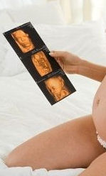
4 D ultrasound is an advanced spatial modelling generated by composite imaging by ultrasound of the fetus. The computer three demensional modelling creates a life-like spatial model of the fetus. 4 D ultrasound is best performed around 28 - 32 weeks. This study is usually not covered by any insurance program and is not billable to the insurance company. The three dimenional images of the fetus in the womb is These unique studies are cash or credit card only ultrasound studies.
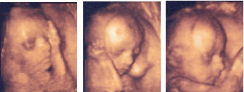
4 D ultrasound of Bella. (She is now one of 15 grandchildren of Dr. and Mrs. James Purdy.) This was taken very early at 25 weeks of pregnancy at Rush Hospital 4 D ultrasound unit.
Ultrasound scanning can be performed by a hand held transducer apparatus. The transducer can image from an external contact on the abdomen or exteranl genitalia (translabial approach or transvaginal approach with the vagina (by vaginal probe insertion) to better image the cervix, uterus and ovaries/fallopian tubes
What are inherent benefits and risk of Ultrasound?
Beneficial values:
- Ultrasound scanning is noninvasive - contact scanning on the body surface
- Painless unless the area scanned is already painful
- Ultrasound is widely used in the medical oficce like Total Care for Women
- easy-to-use
- Less expensive than radiologic imaging eg CT scanning or MRI scanning
- Ultrasound imaging uses no x-radiation waves - only high frequency (non audible ultrasound) sound waves.
- Ultrasound scanning gives a clear picture of soft tissues that do not show up well on x-ray images.
- Ultrasound causes no health problems and may be repeated as often as is necessary.- the energy of the sound waves will not cause tissue harm
- Ultrasound is the preferred imaging method for the diagnosis and monitoring of pregnant women and their unborn babies. (No risk on repeated use)
- Ultrasound has been used to evaluate pregancy develpopment for nearly five decades and there has been no evidence of harm to the patient, embryo or fetus. Nevertheless, ultrasound should be performed only when clinically indicated since there is a cost that insurance companies may not pay if improper indications for use are used.
- Ultrasound allows the doctor to see inside the uterus and provides much information about the pregnancy - Dr. Purdy has always called this ultrasound ".....the window into the womb"as early as the gestational sac size (age).
Risks
- None
What factors limit the use Ultrasound?
- Obesity: deeper tissues require more energy and higher frequency of sound waves to penetrate and image the target organs
- excessive motion may blur the tissue interfaces
- gas in the intestines can cause artifact and distort the image
- image is skill dependent on the sonographer as well as dependent on the machine's processing power - newer machines work faster
- with better images!
Who interprets the results?
A local radiologist (physician trained to supervise and interpret radiology examination and ultrasonography) will evaluate the images and send a signed report to Dr.Purdy. Then Dr. Purdy will share the results with the patiemt. In some cases the sonographer with Dr. Purdy may discuss results with you at the conclusion of your examination such as in SIS procedures or OB ultrasound procedures performed at Total Care for Women - eg fetal positioning or cervical length. If the study is performed a Rush Hospital, then the Rush radiologist(s) will report; if the imaging is performed by Dr. Puppa, Dr. Puppa will report by fax. Other scanning locations (Total Care for Women) will send the report to Meridian Imaging.and then forward the report by fax to Dr. Purdy. All reports will be incorporated into the electroinic medical record system by Greenway at Total Care for Women..
Obstetrical ultrasound: The window into the womb

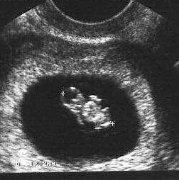
Obstetrical ultrasound can be used to::
- evaluate for single or multiple pregnancies
- evaluate the viability of an early pregnancy by means of the heart rate.
- evaluate by normograms or biometry tables the age of the pregnancy
- evaluate intrauterine anomalies of the growing body parts of the fetus.
- evaluate the fetal position ( vertex, breech, transverse, oblique etc)
- evaluate the location of the placenta and its condition - check for placenta previa (position) or condition ( hemorrhage - abruptio placentae etc) or aging of the placenta
- evalaute the volume of amniotic fluid around the baby - called the AFI (amniotic fluid index)
- evalaute by Doppler the blood flow in the fetus and placental bloos vessels - umbilical arteries and umbical vein
- evaluate cervical length in pregnancy and warn of shortening of the cervical length (below 3.0 cm) as a potential risk factor for preterm labor.
- evalaute fetal growth parameters to monitor the health of the growing fetus.
Gynecologic ultrasound creates (as does Ob ultrasound studies) computer pictures of the body parts of the pelvis including
- boney pelvis
- bladder and urethra
- adnexae (both ovareis and fallopian tubes)
- uterus and cervix and vagina
- rectum
- pelvic blood vessels and pelvic lymph nodes
Gyn ultrasound can be used to:
- evaluate abnormal uterine bleeding (AUB) symptoms
- evaluate causes of pelvic pain, painful periods (dysmenorrhea)
- esvaluate pelvic masses diagnosed on clinical pelvic exams
- evaluate size, shape and growth of fibroid(s) (leiomyoma(s))
- evaluate growth of benignand canceropus tumots of the pelvis
- evaluate the location and size of abcess or hermatoma formation(s)
- evalaute the volume and internal strucutres of ovarian cyst(s)
- evalaute by Doppler the blood flow in the pelvic organs
- evaluate lower abdominal and pelvic anatomy
Saline Sonohysterography (called SIS procedure)
Saline infusion sonohysterography or SIS, is a minimally invasive (MIS) ultrasound procedure tha tis routinely performed in the office setting at Total Care for Women. This procedure images the cervix and the endometrial cavity of the uterus. The saline expands the endometrial cavity for better visualization of the endometrial cavity. It is a safe and essentailly minimally uncomfortable procedure to evaluate the condition of the endometrial cavity.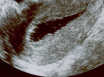
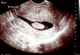
Normal SIS scan
 Abnormal SIS scan with polyp
Saline Sonohysterography can check for cause of abnormal bleeding:
- polyp(s)
- Endometrial mass or submucus fibroid(s)
- Endometrial thickening as found in endometrial overgrowth (hyperplasia)
- atrophy or postmenopausal thickness (less than 4 mm)
- Congenital anomalies: Double endometrial cavity (congenital) or a partial septum (wall)
This procedure can be combined with an endometrial biopsy. This office procedure can replace the more expensive D&C/Hysterocopy in selected cases as an office based minimally invasive procedure (MIS). The procedure (SIS) takes only a few minutes to complete in the office and the discomfort is minimalized by a small warmed saline injection into the cevix and uterine cavity to outline the cavity by ultrasound. Dr. Purdy and the patient see the imaging immediately. The discomfort is less than a menstrual cramp with the use of analgesic medication (Toradol).
How should I prepare for a SIS procedure?
- Clothing: Same type of garments as for a routine pelvic examination. The lower abdomen and external genitalia need to be easily undrapped
- Avoid large solid meals for 6 to 12 hours before the scan in order to avoid unnecesary echo returns from the small and large intestines
- Avoid drinking carbonated or fizzy liquids before the procedure (gas bubbles from sodas will cause too many false echo returns)
- Empty the urinary bladder just before the exam since this procedure requires an emtpy bladder to scan the upper genitalia
- Your modesty will be protected and the technician will be a feamle!
- Same type of garments as for a routine pelvic examination. The lower abdomen and external genitalia need to be easily undrapped
- The patinet lies on her back with her feet in stirrups (as on a gynecology examining table). A speculum is placed in the vagina
- The vagina is washed with either a betadine swab or Hiboclens swab to reduce the risk of an infection after the procedure
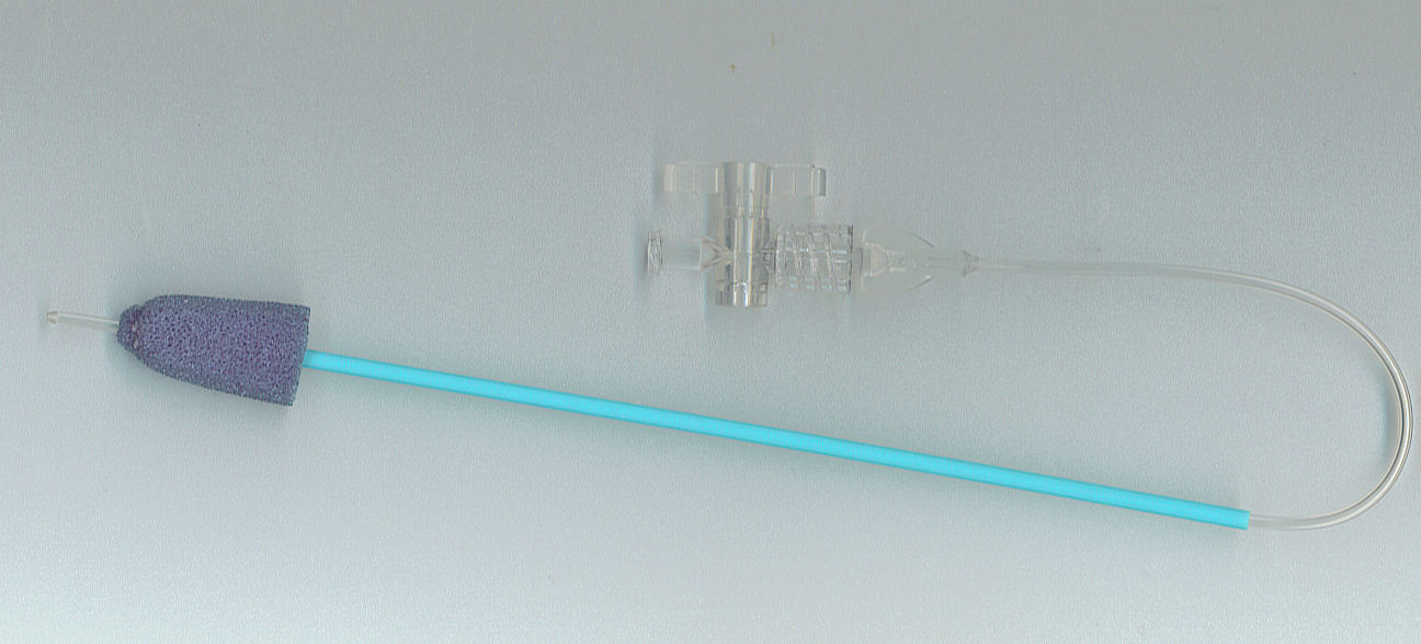
- Next a mall tube with a sponge compressible tip (to create cervical seal) is used to
insert a saline solution into the uterus - less than 3 cc's of solution.
- A vaginal ultrasound probe wrapped in a large condom is inserted to make images of the saline filling the endometrial cavity.
How is the procedure performed?
How uncomfortable is the procedure? The test may cause some discomfort, such as pelvic or lower abdominal cramping.
- Dr. Purdy may recommend a non steroidal mediation such as Motrin or Aleve before the patient comes to the office
- Dr. Purdy will usually recommend Toroadol 30 or 60 mg injection just before or after the procedure for control of some cramps (period like cramps)
- Motrin, Alevel or Cataflam (perscribed by Dr. Purdy) is offered if needed psost SIS procedure for mild cramps that may persistt for a few minutes or 1 -2 hours after the procedure
- Most women can drive and return to work within two hours after the SIS. There are no specific restrictions on activity after the SIS has been done
Post procedure advise:
Contact the nursing staff or Dr. Purdy after the procedure if any of the following symptoms develop:� Persistent pelvic or abdominal pain
� Difficult or painful urination
� Temperature over 100 degrees
How should I prepare for an abdominal or pelvic ultrasound?
- Clothing: Same type of garments as for a routine pelvic examination. The lower abdomen and external genitalia need to be easily undrapped
- Avoid large solid meals for 6 to 12 hours before the scan in order to avoid unnecesary echo returns from the small and large intestines - avoid gas bubbles created by normal digestion
- Unless otherwise directed, drink plenty of fluids such as water to insure a full bladder for the procedure
- Avoid drinking carbonated or fizzy liquids before the procedure (gas bubbles from sodas will cause too many false echo returns)
- Avoid urinating just before the ultrasound scanning procedure - a full bladder is needed for a good pelvic ultrasound
- Your modesty will be protected and the technician will be a feamle!
How should I prepare for a transvaginal ultrasound?
- Clothing: Same type of garments as for a routine pelvic examination. The lower abdomen and external genitalia need to be easily undrapped
- Avoid large solid meals for 6 to 12 hours before the scan in order to avoid unnecesary echo returns from the small and large intestines - - avoid gas bubbles created by normal digestion
- Avoid drinking carbonated or fizzy liquids before the procedure (gas bubbles from sodas will cause too many false echo returns)
- Empty the urinary bladder just before the exam since this procedure requires an emtpy bladder to scan the upper genitalia
- A vaginal probe ultrasound will be used; the insertion will be with a large condom that may feel weird but this insertion should not cause any unnecassry discomfort.
- Your modesty will be protected and the technician will be a feamle!
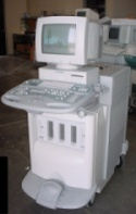
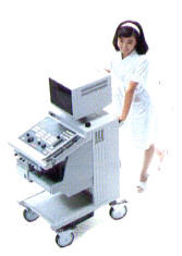
What does the equipment look like?
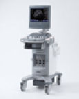 Ultrasound unit like Acuson consist of a console containing a high speed computer that images the reflected ultra high frequency sound generator, a video display screen and a ultrasound transducer that is used to both transmit and receive the the sound waves. The transducer is hand held and can scan the abdomen or the pelvis. The machime transmits varois ultrasound frequency depending on the transducer head array. The higher the frequency, the deeper the sound can penetrate. nner by a cord. IN addition to a hand held traqnsducer, there is a thinner vaginal probe transducer that is long and narrow and it is able to be placed into the vagina to scan the upper genital structures such as the ovaries, fallopian tubes and uterus/endometrium (uterine lining) or posterior back of the pelvis/rectum.
. The ultrasound image is immediately visible on a nearby screen that
looks much like a computer or television monitor. The image is created
based on the amplitude (strength), frequency and time it takes for the
sound signal to return from the patient to the transducer. The portable ultyrasound unit stores the image in computer memory or burns the information to a disc, flash drive or sends it intot he local internetwork for additional viewing elsewhere (virtual radiology).
The ultrasound image is immediately visible on a nearby screen that
looks much like a computer or television monitor. The image is created
based on the amplitude (strength), frequency and time it takes for the
sound signal to return from the patient to the transducer. The portable ultyrasound unit stores the image in computer memory or burns the information to a disc, flash drive or sends it intot he local internetwork for additional viewing elsewhere (virtual radiology).
How does the procedure work?
![]()
The final image that we see is the composite of reeived ultrasound waves (yes - sound waves) that are reflected back from the patient's tissue. This the same principle as the naval sonar device or the imaging of fish that the fisherman down the street uses to find his fish in the water. The advanced circuits in the processing unit receives the reflected high frequency sound waves ( too high to hear - hence ultra high frequency sound). The intensity (amplitude), wave direction and transit time determine the size and distance from the reflecting object (tissue planes). The computer uses all the point sof received sound infomration to create pixels that in turn create an image. The iamges when sequenced at rates exceeding 16 frames a second create for us a moving and living image of our bodies. Essentialy, the calcualtions, pixel development is so fast that we perceive the images in real time. The computer can then compare, measure and colorize to create the image, moving images or plot the blood flow velocity. Various formulas of software make the millions of calculations per second to present the data in an image our eyes can transmto to our brains for the mental image we subsequenbtly see! This truly future science concepy is now a everyday reality.- not science fiction of yesteryears. .
In medicine, ultrasound is used to detect changes in appearance of organs, tissues, and vessels or detect abnormal masses, such as tumors.
How is the procedure performed?
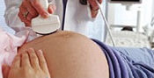
For most ultrasound exams, the patient is positioned is flat on an exam table. At Total Care for Women, the ultrasound room has a special exam table that change into many orientations for ease of examination and patient comfort.
A clear water-based gel is applied to the area of the body being studied to help the transducer make secure contact with the body and eliminate air pockets between the transducer and the skin. The sonographer then presses the transducer firmly against the skin and sweeps it over the area of interest.
Transvaginal scan needs to be performed. This technique often provides improved, more detailed images of the uterus and ovaries. This method of scanning is especially useful in early pregnancy.
Transv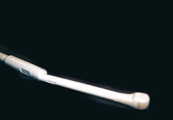 aginal ultrasound is performed very much like a gynecologic exam and involves the insertion of the transducer into the vagina after the patient empties her bladder. The tip of the transducer is smaller than the standard speculum used when performing a pap smear.
A protective cover (large condom) is placed over the transducer, lubricated with a
small amount of gel, and then inserted into the vagina.
aginal ultrasound is performed very much like a gynecologic exam and involves the insertion of the transducer into the vagina after the patient empties her bladder. The tip of the transducer is smaller than the standard speculum used when performing a pap smear.
A protective cover (large condom) is placed over the transducer, lubricated with a
small amount of gel, and then inserted into the vagina.
 What will I experience during and after the procedure?
After you are positioned on the examination table, the female sonographer at Total CAre for Women will apply some warm water-based gel on your skin and then place the transducer firmly against your body, moving it back and forth over the area of interest until the desired images are captured. The gel will cool and the skin may feel cooler or cold. This is normal. There is no pain with the external examination. The vaginal probe transducer is covered with a large condom before insertion into the vagina. The insertion process should not cause any real pain. It amy feel weird but not pauinfull.
If scanning is performed over an area of tenderness, you may feel pressure or minor pain from the transducer. If an area is pathologic, the area scanned may be uncomfortable to touch. The less probe pressure, the less discomfort. The sonographer varies the pressure of the transducer to reduce any discomfort. The actual ultrasound scanning will not cause any pain.
Once the imaging is complete, the gel will be wiped off your skin.
After an ultrasound exam, you should be able to resume your normal with no down time. This a minimally invasive procedure with no real pain or risks to the patien tor if she is pregnant there is no to the embryo or fetus..